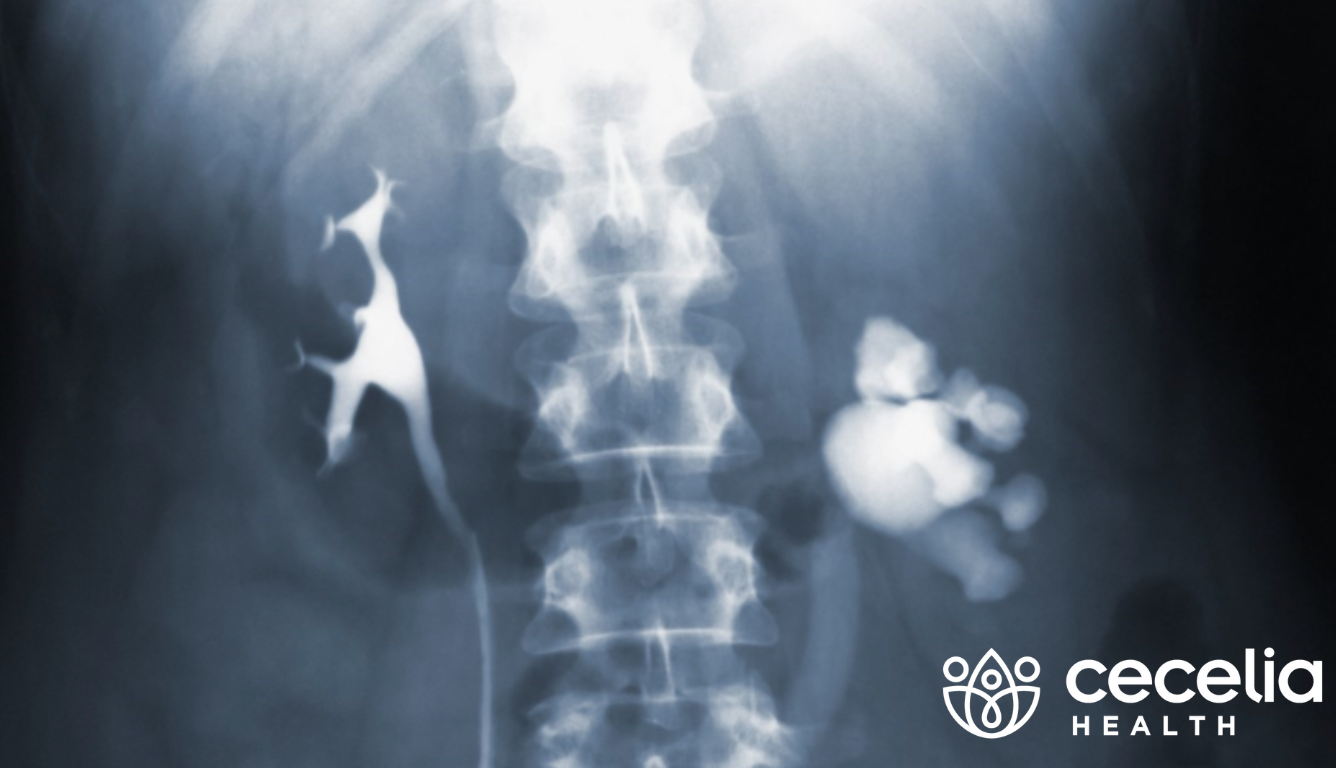
Early stages of Chronic Kidney Disease (CKD) may have very mild or undetectable signs and symptoms even though damage has begun to occur. A blood test can reveal the filtration capacity of the kidneys in the form of an estimated glomular filtration rate (eGFR) lab test. The eGFR can help determine how efficiently the kidneys are currently functioning. Whereas, physical damage is seen with imaging such as renal ultrasound, CT scan, MRI, and kidney biopsy analysis. But which tests are best to determine damage in Chronic Kidney Disease? Let’s take a look at the pros and cons of various imaging techniques.
Detecting Kidney Damage
Real Ultrasound
Real Ultrasound is a testing method that uses sound waves to make images of the kidneys, ureters, and bladder. Black and white computer-generated images are used to see the internal structure of the kidneys to detect kidney cancer, inflammation, and chronic kidney disease.
- Pros – the most commonly used test to identify issues with the kidneys because it can look at the kidney size and internal echo characteristics while providing a lower emittance of radiation.
- Cons – the presence of gas in the abdomen may affect image quality and sometimes the kidneys may appear normal even in kidney disease.
MRI
MRI consists of two types of magnetic resonance imaging (MRI) scans for the kidneys: imaging with or without contrast agents. The contrast agent is a dye or liquid injected into the body to make soft tissues more visible during the imaging process. Newer contrast agents containing gadolinium are safer for kidney patients.
- Pros – can be used in patients with known contraindications to contract mediums or dyes. In addition, growths or carcinomas are more easily detected. In addition, auto contrast MRI can show infiltrative damage through cystic structures to clearly see the blood vessels and lymph nodes to find and treat kidney disease early.
- Cons – contrast agents and dyes are not ideal for the kidneys as it takes longer to eliminate the waste from the body. In addition, like other types of imaging, repeated exposure to radiation may have adverse effects.
CT scan
CT scan is a computed tomography (CT) imaging method that can provide more detailed information about the kidneys than a standard X-ray image via cross-sectional images.
- Pros – a CT scan is often the preferred test to detect renal infections as it can identify gas, stones, calcifications as well as bleeding, abscesses, and obstructions.
- Cons – in some cases, contrast dyes can be contraindicated due to other medical conditions or may cause direct damage, especially in patients who take certain medications. This type of imaging also exposes people to radiation and may be recommended as a secondary test.
Kidney Biopsy
Kidney Biopsy is a procedure to remove a small piece of kidney tissue that can be examined under a microscope for signs of damage or disease.
Pros – this type of imaging can be used when a direct cause cannot be adequately predicted by other imaging procedures. Although invasive, this technique does not expose the body to any radiation and may assist in determining a specific pathological diagnosis for ongoing CKD related symptoms.
Cons – possible risks include bleeding or blood in the urine and pain at the biopsy site. It also has the potential for developing an abnormal connection between two blood vessels called a fistula.
For general CKD damage assessment, renal ultrasound provides a convenient method to view the size and shape of the kidneys to indicate disease progression. If further imaging is needed to detect specific abnormalities, MRI is often used clinically to assess carcinoma or other masses. A CT scan is useful to detect tumors, lesions or obstructive conditions of the kidneys. Whereas a kidney biopsy provides imaging of specific tissues to resolve pathology issues.
Discuss the options with your kidney doctor or health care provider based on personal medical history risks and medication use. Take into account the length and frequency expected for imaging tests to determine risks of radiation exposure and ask about the type of equipment as well as the age and safety. Technology in the field of medicine has provided lifesaving opportunities for detection and will continue to evolve over time.